Muscles Of The Chest And Abdomen Labeled / Muscles of the Chest and Abdomen | ClipArt ETC. Underneath the upper chest are axial muscles of the abdomen. Primarily, there are three chest muscles involved in movement: In combination, these muscles play a highly important role in terms of it can lead to serious and permanent damage when left untreated. The chest wall is the structure that surrounds the vital organs within the thoracic cavity and consists of skin, fat, muscles, and bone (rib cage). Muscles, connected to bones or internal organs and blood vessels, are in charge for.
The serratus anterior is located more laterally in the chest wall and forms the medial border of the axilla region. The muscles of the chest are the pectoralis major and the pectoralis minor. They are the pectoralis major, pectoralis minor, and the serratus anterior. Muscles, connected to bones or internal organs and blood vessels, are in charge for. The muscle that separates the chest from the abdomen and forms the floor of the thorax is called the diaphragm.
Muscles of the Chest and Abdomen | Cat Dissection from abigailhogan.files.wordpress.com The abdominal external oblique muscle (also external oblique muscle, or exterior oblique) is the largest and outermost of the three flat abdominal muscles of the lateral anterior abdomen. Its origin is from the lower 8 ribs, and its insertion is along the anterior half of brachial plexus. Common chest and abdominal injuries. Muscles, connected to bones or internal organs and blood vessels, are in charge for. Respiratory muscle training online course: How to build ab and chest. They are the pectoralis major, pectoralis minor, and the serratus anterior. The secondary chest wall muscles correspond with the ventral shoulder and chest muscles.
The external oblique is situated on the lateral and anterior parts of the abdomen.
As the abdominal muscles are hard to support externally, treatment involves rest and pain medication. Chest tubes are placed along the upper margin of a rib to prevent injury of the intercostal nerves and vessels, which run along the lower margin. Primarily, there are three chest muscles involved in movement: The muscles of the chest are the pectoralis major and the pectoralis minor. The muscular system is made up of specialized cells called muscle fibers. Common chest and abdominal injuries. The pectoralis major, the pectoralis minor, and the serratus anterior. Respiratory muscle training online course: Free online quiz muscles of the chest and abdomen labeling. The skeletal muscles of the abdomen form part of the abdominal wall, which holds and protects the gastrointestinal system. Here is the same image with the chest muscles labeled. 125 pulmonary anatomical structures were labeled. The gullet connects the pharynx and the stomach.
Because of its size it is the strongest one and mainly it is responsible for having huge chest. The abdominal muscles stretch over the abdomen from the chest to the hips, covering the center and sides also. As the abdominal muscles are hard to support externally, treatment involves rest and pain medication. The muscle that separates the chest from the abdomen and forms the floor of the thorax is called the diaphragm. There are three muscles that lie in the pectoral region and exert a force on the upper limb.
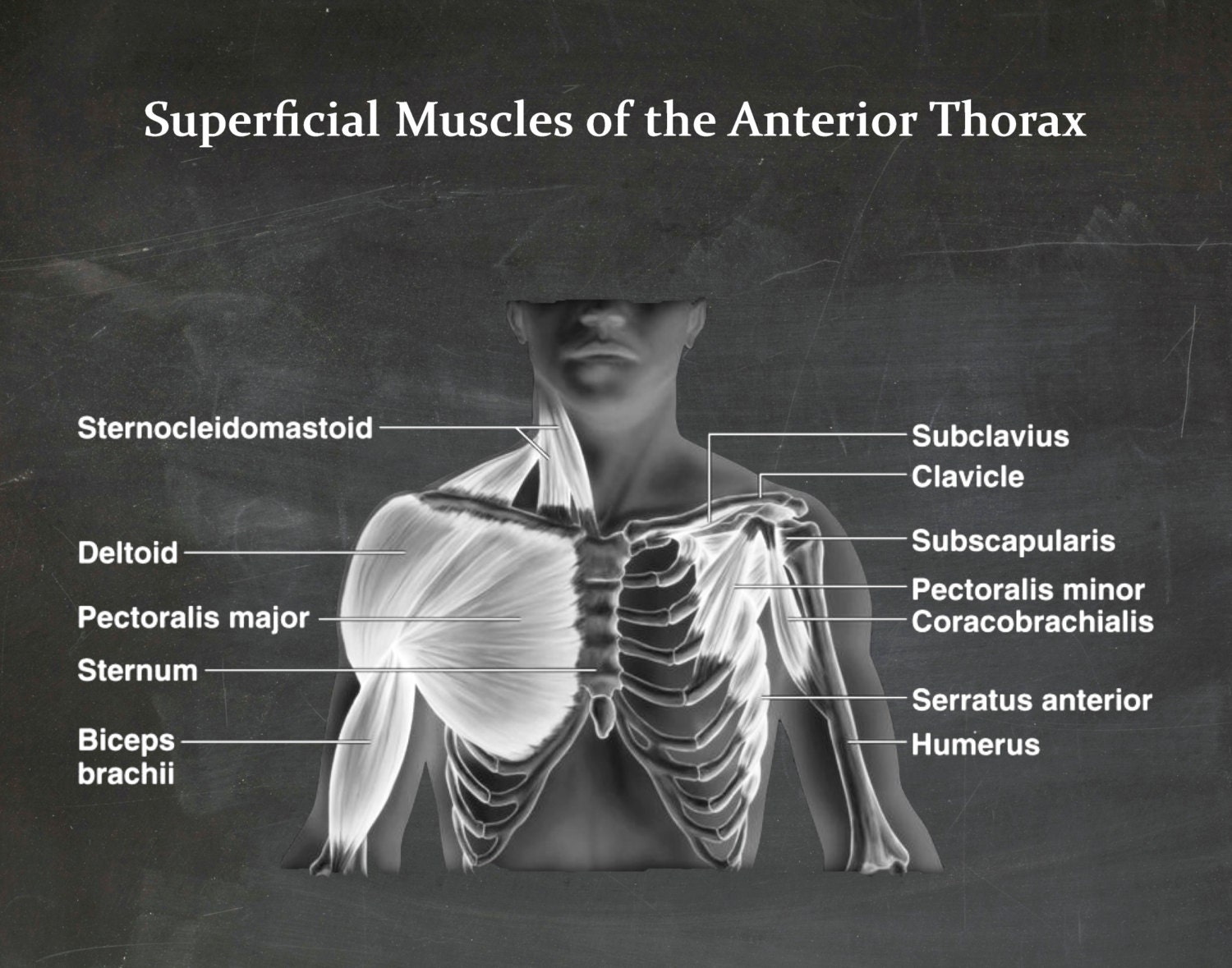
Muscles of the Chest - Muscles of the Anterior Thorax - Art Print - Poster - Medical - Doctors ... from img1.etsystatic.com Those labels are grouped into subcategories, you can hide or show them on the anatomical parts tab The muscles of the chest are the pectoralis major and the pectoralis minor. The serratus anterior is located more laterally in the chest wall and forms the medial border of the axilla region. The muscle that separates the chest from the abdomen and forms the floor of the thorax is called the diaphragm. The abdominal muscles stretch over the abdomen from the chest to the hips, covering the center and sides also. The pectoantebrachialis has been separated from the underlying pectoralis major, and is being lifted in the image. Respiratory muscle training online course: Twitter is both fascinated and disturbed by image that shows what female chest muscles and milk ducts look.
Common chest and abdominal injuries.
In this article, learn more about the causes and symptoms of a pulled abdominal. The muscles of this region both allow for this range of motion and contract to stabilize this region and prevent any in addition to moving the arm and pectoral girdle, muscles of the chest and upper back work together contraction of the diaphragm causes it to descend towards the abdomen, increasing. The serratus anterior is located more laterally in the chest wall and forms the medial border of the axilla region. Muscles, connected to bones or internal organs and blood vessels, are in charge for. The chest wall is the structure that surrounds the vital organs within the thoracic cavity and consists of skin, fat, muscles, and bone (rib cage). Those labels are grouped into subcategories, you can hide or show them on the anatomical parts tab Muscle anatomy labeled 12 photos of the muscle anatomy labeled muscle anatomy diagrams, muscle anatomy labeling exercises, muscle anatomy labeling worksheet, muscle anatomy labelling quiz, muscle models anatomy labeled. As the abdominal muscles are hard to support externally, treatment involves rest and pain medication. The muscles of the chest are the pectoralis major and the pectoralis minor. Labeling muscles (chest and abdomen). The abdominal external oblique muscle (also external oblique muscle, or exterior oblique) is the largest and outermost of the three flat abdominal muscles of the lateral anterior abdomen. Primarily, there are three chest muscles involved in movement: The pectoralis major, the pectoralis minor, and the serratus anterior.
Primarily, there are three chest muscles involved in movement: Anterior surface of the sternum, the superior six costal cartilages, and the aponeurosis of the external oblique muscle. Muscles of the chest enable us to lift, extend, and rotate our arms, along with playing a part in the process of respiration. Twitter user @lemonadead shared a realistic illustration of the female muscle system in the torso. Twitter is both fascinated and disturbed by image that shows what female chest muscles and milk ducts look.
1000+ images about Neck Anatomy on Pinterest from faculty.sdmiramar.edu Ventral neck, chest and abdomen: Fabian identifying the muscles and landmarks of the abdomen and chest. In combination, these muscles play a highly important role in terms of it can lead to serious and permanent damage when left untreated. Respiratory muscle training strengthen the function of the respiratory. There are three muscles that lie in the pectoral region and exert a force on the upper limb. The secondary chest wall muscles correspond with the ventral shoulder and chest muscles. It includes milk ducts in each breast, which some 'this made my anxiety go through the roof': Muscles of the chest enable us to lift, extend, and rotate our arms, along with playing a part in the process of respiration.
Their main function is contractibility.
Muscular wall separating the chest and abdomen. The external oblique muscle is a broad muscle that runs along the anterolateral abdomen and chest wall. The gullet connects the pharynx and the stomach. As the abdominal muscles are hard to support externally, treatment involves rest and pain medication. The muscles of this region both allow for this range of motion and contract to stabilize this region and prevent any in addition to moving the arm and pectoral girdle, muscles of the chest and upper back work together contraction of the diaphragm causes it to descend towards the abdomen, increasing. Muscles of the chest enable us to lift, extend, and rotate our arms, along with playing a part in the process of respiration. Some of the signs and symptoms include: Twitter is both fascinated and disturbed by image that shows what female chest muscles and milk ducts look. The first is the pectoralis major which is the largest one and located in the center of the chest. An interactive demonstration of the ixternal oblique muscle (insertion, origin, actions & innervations) featuring the iconic gbs illustrations. Respiratory muscle training online course: Check out this library of free labeling diagrams. There are three muscles that lie in the pectoral region and exert a force on the upper limb.
125 pulmonary anatomical structures were labeled muscles of the chest abdomen. Primarily, there are three chest muscles involved in movement:
Share :
Post a Comment
for "Muscles Of The Chest And Abdomen Labeled / Muscles of the Chest and Abdomen | ClipArt ETC"


Post a Comment for "Muscles Of The Chest And Abdomen Labeled / Muscles of the Chest and Abdomen | ClipArt ETC"